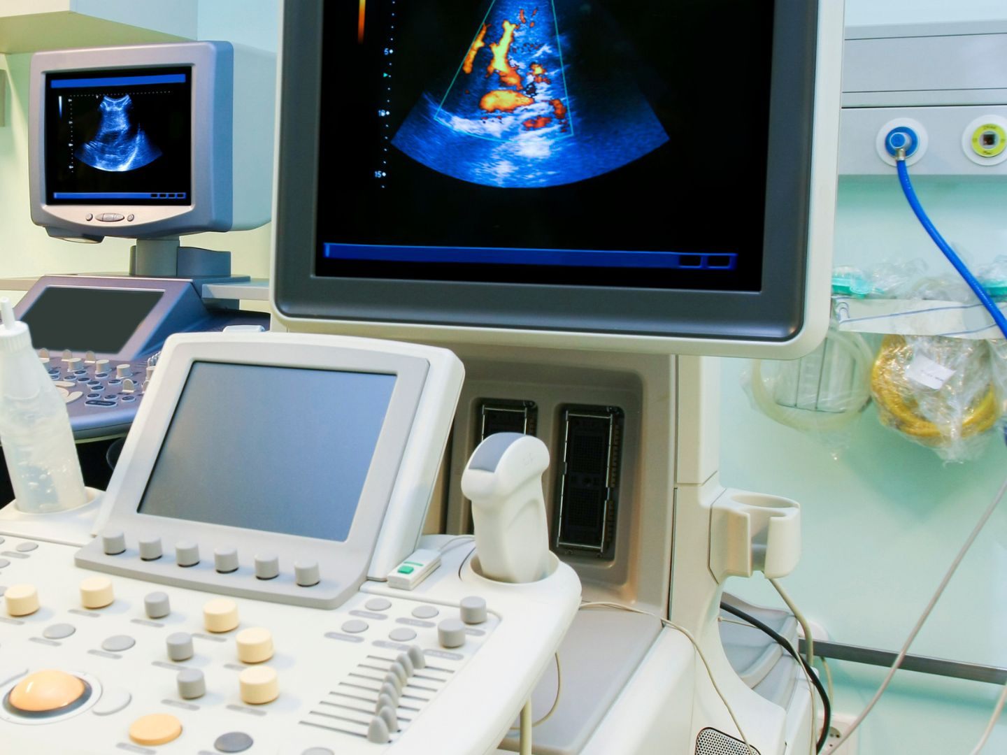
Stroke Diagnosis in Newborns Now Possible via Ultrasound

"Doppler sonography can detect blood flow in the vessels of the brain," said Jörg Jüngert, Deputy Head of the Pediatrics Section of the German Society for Ultrasound in Medicine (DEGUM), at a press conference of the German professional society.
Widespread Use of Sonography
The use of sonography, which operates with sound waves without radiation exposure, has become particularly widespread in pediatrics in recent years. Ultrasound devices are also very often available in private medical practices. For a quick clarification of a suspected illness in babies, this is a great advantage outside of hospitals. Worldwide attention was drawn, for example, to the ultrasound-guided local anesthesia in children after injuries, which was first developed in Vienna.
However, development continues. "In newborns and babies, the entire brain can be depicted using ultrasound. Sonography plays a central role here," said Jüngert. During the ultrasound examination of the brain, doctors take advantage of the fact that after birth, the skull skeleton in infants, with the fontanelles, the tissue gaps on the head, is not yet ossified and thus permeable to ultrasound.
Ultrasound Advancement: MRI No Longer Needed for Babies
The senior physician at the Children's and Youth Clinic of the University Hospital Erlangen pointed out a problem that existed until now: "Tissue perfusion or the smallest vessels with low flow velocity could not always be adequately depicted and characterized with this method."
This has changed recently and also makes it possible to clarify a suspected stroke even in newborns. The expert: "Here, contrast-enhanced sonography can help directly at the bedside."
In itself, a magnetic resonance imaging (MRI) would be the decisive procedure. However, this requires the availability of such devices, is associated with the transport of small babies, and the administration of sedative medications to keep them still during the examination in the devices.
Ultrasound Localization Microscopy
At the clinic in Erlangen, contrast-enhanced sonography was recently used for the first time in a suspected stroke case in a baby. The contrast agent consists of microscopically small gas bubbles with a fat shell, which later dissolve. The harmless gas is then exhaled by the child without any other effects.
"A commercially available ultrasound device with special software makes it possible to visualize the blood flow in the brain in real-time and to characterize the smallest vessels using a special analysis method. This procedure is referred to as Ultrasound Localization Microscopy (ULM)," stated the DEGUM at the press conference on Monday.
Microscopy Sonography Not Yet Approved in Europe
Stroke is generally considered a disease of older people - however, for example, up to 200 newborns in Germany suffer such an acute neurological event annually. There is an increased risk in premature infants. High-resolution sonography is also an important imaging procedure for detecting pathological changes in the brain of premature and newborn infants. A preliminary drawback: In Europe, microscopy sonography with the gas bubble contrast agent is not yet properly approved. In the USA, it was developed, for example, for examining liver blood flow in children.
(APA/Red.)
This article has been automatically translated, read the original article here.


